Michigan Diagnostic Pathologists
Michigan Diagnostic Pathologists, PLLC
Affiliated with: Ascension Providence - Providence Park Hospitals, Southfield, Novi and Rochester Campuses
 
Acute Appendicitis
Acute Appendicitis

Acute appendicitis is inflammation of the appendix, a small pouch that hangs off of the beginning of the large intestine. Thus usually occurs due to an obstruction and can become infected.

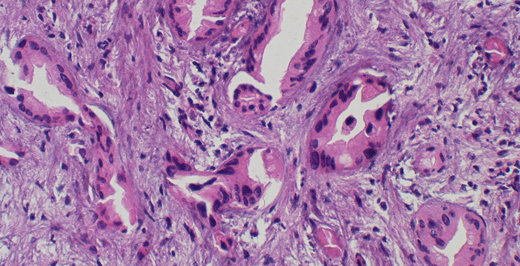
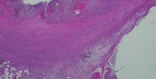
In this image, the various layers of the appendix can be seen. The purplish haze seen through the appendiceal layers is evidence of the inflammatory cells.

For more information, click here

Cholangiocarcinoma
Cholangiocarcinoma

Cholangiocarcinoma is a cancer of the biliary tree; a system of ducts that carries bile from the liver to the small intestine where it is used to aid in digestion.

In image 2, the ducts, which appear as large circular structures, are disorganized with irregular lumens. The nuclei of their cells, which appear as smaller dark purple structures, show variation in shape and size when in normal ducts they should be the same size and shape. In image 3, the ducts are again disorganized. In this image, mucinous secretions are more evident than in image 2.

For more information, click here

Hairy Cell Leukemia
Hairy Cell Leukemia

Hairy cell leukemia is a cancer of a type of white blood cell called the B-cell. Hairy cell leukemia involves the peripheral blood, bone marrow, liver and spleen.

In this image, near the center of the field, are several cells with round, bland nuclei and abundant pale cytoplasm. Sometimes this cytoplasm can have multiple thin projections, hence the name hairy cell leukemia.

For more information, click here

Congo Red Positive Amyloid Deposits in the GI Tract

Amyloid deposits are insoluble collections of misfolded proteins. These misfolded proteins can be involved in a number of disease processes including amyloidosis, Alzheimer’s disease, rheumatoid arthritis and many more.

In this image, the amyloid deposits are highlighted by a special stain called Congo Red which gives them a bright red-orange color under standard microscopy. Under polarized light the amyloid will have an apple-green color.

For more information, click here


BILLING

Pathologists practice independently from your primary care physician or specialist. This is why you may receive a bill from your pathologist in addition to your standard physician bill. If you have received a bill from Michigan Diagnostic Pathologists, your physician has requested us to test your blood, tissue specimen, examine your pap smear or perform some other exam. Our services are billed by pathology billing specialists who forward payment claims to your insurance company.

Our bill will be separately received from those sent by your doctor, hospital or laboratory. Our services are not covered by co-pays made at the time you visited your doctor. Co-payments strictly cover physician visits and do not cover our pathology services.

If you have questions about a bill, feel free to contact our billing service. All billing questions are handled in strict confidence. For general questions about billing, please visit the Contact Us page.

FOR HEALTH CARE PROVIDERS